

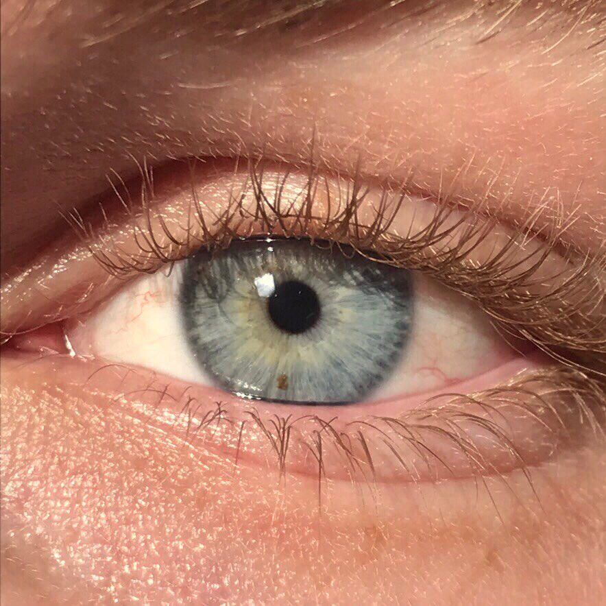
How does the human eye stock the tiny central zone of the retina with exactly the right mix of retinal cones? Researchers at Johns Hopkins University say they have found the chemical instructions — and in the process overturned a long-standing theory about how our sharpest vision is built.
Two Signals Shape How Retinal Cones Form
Working with miniature retinal organoids grown in the laboratory and watched over several months, the team found that two signalling molecules divide the labour. Retinoic acid limits the formation of blue-sensitive cones in the central retina, while thyroid hormones encourage developing cells to become red- or green-sensitive instead. The foveola — the pinpoint region these cells populate — is estimated to contribute around half of human visual perception, so the recipe that builds it is fundamental to how we see.
Transformation, Not Migration
The study challenges the older idea that blue-sensitive cones simply migrate out of the foveola as the eye matures. Instead, the researchers observed blue-sensitive cones appearing in the foveola during weeks 10 to 12 of fetal development, with some of them converting into red- and green-sensitive cones by week 14 — transforming in place under hormonal direction. Follow more science and health discoveries for more research news of this kind.
Why It Matters Beyond the Lab
Understanding the developmental signals could eventually help scientists grow replacement photoreceptors for conditions such as macular degeneration and glaucoma, where cone loss steals central vision. That is a long road — organoid insights must still be translated into therapies — but as the detailed report on the discovery explains, knowing which switches control cone identity is a prerequisite for any attempt to manufacture the right cells.
It is a reminder that even in an organ as studied as the eye, basic developmental biology still holds surprises — and that some of them may one day help restore sight.
